The Application of 3D Printing Technology in Otological Diagnosis and Treatment from Morphological Reconstruction to Functional Restoration
DOI:
https://doi.org/10.71321/mgsr6v33Keywords:
3D printing, otology, morphological reconstruction, functional restoration, personalized medicineAbstract
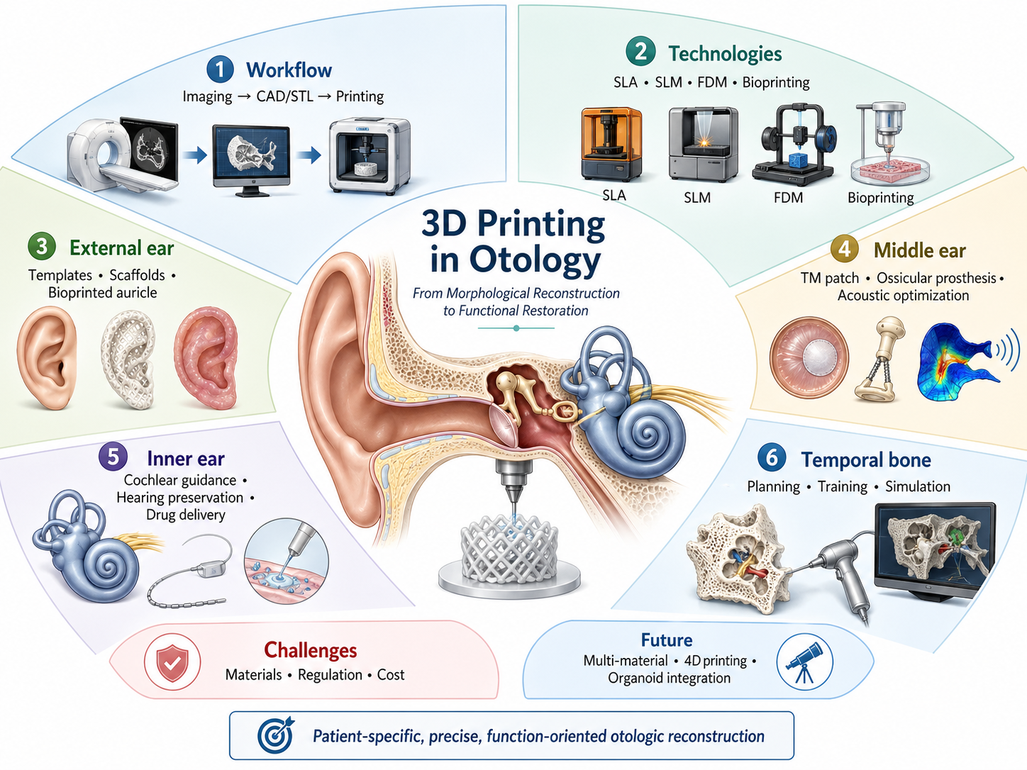
Through patient-specific design and precise anatomical fabrication, 3D printing is reshaping otology from experience-based surgery toward digital precision medicine. This review summarizes recent advances in the application of 3D printing in otology, focusing on technical platforms, morphological reconstruction, and functional restoration. At the technical level, stereolithography, selective laser melting, fused deposition modeling, and 3D bioprinting are suited to preoperative simulation, titanium-alloy implant fabrication, educational models, and tissue-engineered scaffolds, respectively. For morphological reconstruction, 3D printing enables precise replication of structures ranging from auricular frameworks and middle-ear ossicular chains to temporal-bone anatomical models. For functional restoration, patient-specific ossicular prostheses can optimize sound conduction, surgical guides can assist cochlear implantation and help preserve residual hearing, sustained-release devices may help overcome barriers to inner-ear drug delivery, and tissue-engineered scaffolds have opened new possibilities for inner-ear neural repair and regeneration. Although challenges related to material compatibility, regulatory approval, and cost-effectiveness remain, advances in multi-material printing and 4D printing may further expand the clinical potential of 3D printing in personalized otologic reconstruction that integrates anatomical form with functional recovery.
References
[1] M. Stavrakas et al., ‘3D Printing in ENT Training: Current State-of-the-Art and Perspectives’, Maedica, vol. 19, no. 3, Sep. 2024, doi: 10.26574/maedica.2024.19.3.629.
[2] F. Zoccali et al., ‘3D Printing in Otolaryngology Surgery: Descriptive Review of Literature to Define the State of the Art’, Healthcare, vol. 11, no. 1, p. 108, Dec. 2022, doi: 10.3390/healthcare11010108.
[3] T. Kaneko, ‘A System for Three-Dimensional Shape Measurement and its Application in Microtia Ear Reconstruction.’, Keio j. med, vol. 42, no. 1, pp. 22–40, 1993, doi: 10.2302/kjm.42.22.
[4] N. Zhong and X. Zhao, ‘3D printing for clinical application in otorhinolaryngology’, Eur Arch Otorhinolaryngol, vol. 274, no. 12, pp. 4079–4089, Dec. 2017, doi: 10.1007/s00405-017-4743-0.
[5] M. A. De Souza, R. F. Bento, P. T. Lopes, D. M. De Pinto Rangel, and L. Formighieri, ‘Three-dimensional printing in otolaryngology education: a systematic review’, Eur Arch Otorhinolaryngol, vol. 279, no. 4, pp. 1709–1719, Apr. 2022, doi: 10.1007/s00405-021-07088-7.
[6] L. F. Velásquez-García and Y. Kornbluth, ‘Biomedical Applications of Metal 3D Printing’, Annu. Rev. Biomed. Eng., vol. 23, no. 1, pp. 307–338, Jul. 2021, doi: 10.1146/annurev-bioeng-082020-032402.
[7] M. Larobina, ‘Thirty Years of the DICOM Standard’, Tomography, vol. 9, no. 5, pp. 1829–1838, Oct. 2023, doi: 10.3390/tomography9050145.
[8] B. C. Gross, J. L. Erkal, S. Y. Lockwood, C. Chen, and D. M. Spence, ‘Evaluation of 3D Printing and Its Potential Impact on Biotechnology and the Chemical Sciences’, Anal. Chem., vol. 86, no. 7, pp. 3240–3253, Apr. 2014, doi: 10.1021/ac403397r.
[9] M. Pérez-Sevilla, F. Rivas-Navazo, P. Latorre-Carmona, and D. Fernández-Zoppino, ‘Protocol for Converting DICOM Files to STL Models Using 3D Slicer and Ultimaker Cura’, JPM, vol. 15, no. 3, p. 118, Mar. 2025, doi: 10.3390/jpm15030118.
[10] A. Marro, T. Bandukwala, and W. Mak, ‘Three-Dimensional Printing and Medical Imaging: A Review of the Methods and Applications’, Current Problems in Diagnostic Radiology, vol. 45, no. 1, pp. 2–9, Jan. 2016, doi: 10.1067/j.cpradiol.2015.07.009.
[11] A. Omari, M. Frendø, M. S. Sørensen, S. A. W. Andersen, and A. Frithioff, ‘The cutting edge of customized surgery: 3D-printed models for patient-specific interventions in otology and auricular management—a systematic review’, Eur Arch Otorhinolaryngol, vol. 279, no. 7, pp. 3269–3288, Jul. 2022, doi: 10.1007/s00405-022-07291-0.
[12] A. G. Micco, ‘Are Simulated or 3D Printed Temporal Bones as Useful for Training as Cadaveric Bones’, Laryngoscope, vol. 133, no. 8, pp. 1782–1783, Aug. 2023, doi: 10.1002/lary.30704.
[13] C. L. Reighard, S. J. Hollister, and D. A. Zopf, ‘Auricular reconstruction from rib to 3D printing’, J 3D Print Med, vol. 2, no. 1, pp. 35–41, Jan. 2018, doi: 10.2217/3dp-2017-0017.
[14] A. Frithioff, M. Frendø, D. B. Pedersen, M. S. Sørensen, and S. A. Wuyts Andersen, ‘3D‐Printed Models for Temporal Bone Surgical Training: A Systematic Review’, Otolaryngol.--head neck surg., vol. 165, no. 5, pp. 617–625, Nov. 2021, doi: 10.1177/0194599821993384.
[15] G. Iannella et al., ‘The SAPIENS 3D-printed temporal bone model: a real tool for advanced otologic surgery education’, Eur Arch Otorhinolaryngol, vol. 282, no. 6, pp. 3051–3063, Jun. 2025, doi: 10.1007/s00405-024-09199-3.
[16] K. Takahashi et al., ‘Creating an Optimal 3D Printed Model for Temporal Bone Dissection Training’, Ann Otol Rhinol Laryngol, vol. 126, no. 7, pp. 530–536, Jul. 2017, doi: 10.1177/0003489417705395.
[17] L. M. Markodimitraki, T. C. Ten Harkel, R. L. A. W. Bleys, I. Stegeman, and H. G. X. M. Thomeer, ‘Cochlear implant positioning and fixation using 3D-printed patient specific surgical guides; a cadaveric study’, PLoS ONE, vol. 17, no. 7, p. e0270517, Jul. 2022, doi: 10.1371/journal.pone.0270517.
[18] A. Torrisi et al., ‘The Promise and Challenges of 3D Bioprinting in Otolaryngology: A Contemporary Perspective Viewpoint’, Clinical Otolaryngology, vol. 50, no. 5, pp. 795–807, Sep. 2025, doi: 10.1111/coa.14333.
[19] Y. Huang et al., ‘The application and progress of tissue engineering and biomaterial scaffolds for total auricular reconstruction in microtia’, Front. Bioeng. Biotechnol., vol. 11, p. 1089031, Sep. 2023, doi: 10.3389/fbioe.2023.1089031.
[20] S.-W. Wu, Z.-Z. Nian, W. Lin, and X.-D. Zhang, ‘Unveiling the Intricacies of the Inner Ear Anatomy: Novel 3D-Printed Model for Detailed Visualization and Functional Demonstrations’, J. Laryngol. Otol., vol. 138, no. 7, pp. 787–791, Jul. 2024, doi: 10.1017/S0022215124000367.
[21] W. Zhang et al., ‘Functional hydrogel empowering 3D printing titanium alloys’, Materials Today Bio, vol. 30, p. 101422, Feb. 2025, doi: 10.1016/j.mtbio.2024.101422.
[22] A. McMillan, N. McMillan, N. Gupta, S. P. Kanotra, and A. K. Salem, ‘3D Bioprinting in Otolaryngology: A Review’, Adv Healthcare Materials, vol. 12, no. 19, p. 2203268, Jul. 2023, doi: 10.1002/adhm.202203268.
[23] N. Hirano et al., ‘Ethanol treatment of nanoPGA/PCL composite scaffolds enhances human chondrocyte development in the cellular microenvironment of tissue-engineered auricle constructs’, PLOS ONE, vol. 16, no. 7, p. e0253149, Jul. 2021, doi: 10.1371/journal.pone.0253149.
[24] T. D. Crafts, S. E. Ellsperman, T. J. Wannemuehler, T. D. Bellicchi, T. Z. Shipchandler, and A. V. Mantravadi, ‘Three-Dimensional Printing and Its Applications in Otorhinolaryngology-Head and Neck Surgery’, Otolaryngol Head Neck Surg, vol. 156, no. 6, pp. 999–1010, Jun. 2017, doi: 10.1177/0194599816678372.
[25] D. V. Luquetti, C. L. Heike, A. V. Hing, M. L. Cunningham, and T. C. Cox, ‘Microtia: epidemiology and genetics’, Am J Med Genet A, vol. 158A, no. 1, pp. 124–139, Jan. 2012, doi: 10.1002/ajmg.a.34352.
[26] Y. Guo et al., ‘Innovative tissue engineering strategies for auricular regeneration in microtia: Current advances and future perspectives’, Regenerative Therapy, vol. 30, pp. 665–680, Dec. 2025, doi: 10.1016/j.reth.2025.08.010.
[27] R. C. Tanzer, ‘TOTAL RECONSTRUCTION OF THE EXTERNAL EAR’:, Plastic and Reconstructive Surgery, vol. 23, no. 1, pp. 1–15, Jan. 1959, doi: 10.1097/00006534-195901000-00001.
[28] J. A. Cabin, M. Bassiri-Tehrani, A. P. Sclafani, and T. Romo, ‘Microtia Reconstruction’, Facial Plastic Surgery Clinics of North America, vol. 22, no. 4, pp. 623–638, Nov. 2014, doi: 10.1016/j.fsc.2014.07.004.
[29] R. Cenzi, A. Farina, L. Zuccarino, and F. Carinci, ‘Clinical outcome of 285 Medpor grafts used for craniofacial reconstruction’, J Craniofac Surg, vol. 16, no. 4, pp. 526–530, Jul. 2005, doi: 10.1097/01.scs.0000168761.46700.dc.
[30] J. D. Williams, T. Romo, A. P. Sclafani, and H. Cho, ‘Porous High-Density Polyethylene Implants in Auricular Reconstruction’, Archives of Otolaryngology - Head and Neck Surgery, vol. 123, no. 6, pp. 578–583, Jun. 1997, doi: 10.1001/archotol.1997.01900060020003.
[31] J. Y. Park, Y.-J. Choi, J.-H. Shim, J. H. Park, and D.-W. Cho, ‘Development of a 3D cell printed structure as an alternative to autologs cartilage for auricular reconstruction’, J Biomed Mater Res B Appl Biomater, vol. 105, no. 5, pp. 1016–1028, Jul. 2017, doi: 10.1002/jbm.b.33639.
[32] H.-Y. Chen, L.-S. Ng, C.-S. Chang, T.-C. Lu, N.-H. Chen, and Z.-C. Chen, ‘Pursuing Mirror Image Reconstruction in Unilateral Microtia: Customizing Auricular Framework by Application of Three-Dimensional Imaging and Three-Dimensional Printing’, Plast Reconstr Surg, vol. 139, no. 6, pp. 1433–1443, Jun. 2017, doi: 10.1097/PRS.0000000000003374.
[33] Y. Deng et al., ‘Three-dimensional printing technology for the positioning of the reconstructed auricle in microtia reconstruction’, Journal of Stomatology Oral and Maxillofacial Surgery, vol. 126, no. 4, p. 102207, Sep. 2025, doi: 10.1016/j.jormas.2024.102207.
[34] S. Gao et al., ‘3D printing tissue-engineered scaffolds for auricular reconstruction’, Mater Today Bio, vol. 27, p. 101141, Jul. 2024, doi: 10.1016/j.mtbio.2024.101141.
[35] H. Wang et al., ‘Chondrocyte-laden gelatin/sodium alginate hydrogel integrating 3D printed PU scaffold for auricular cartilage reconstruction’, International Journal of Biological Macromolecules, vol. 253, p. 126294, Dec. 2023, doi: 10.1016/j.ijbiomac.2023.126294.
[36] P. Fisch et al., ‘Tissue Engineered Human Elastic Cartilage From Primary Auricular Chondrocytes for Ear Reconstruction’, Advanced Functional Materials, vol. n/a, no. n/a, p. e30253, doi: 10.1002/adfm.202530253.
[37] M. Mohseni-Dargah, C. Pastras, P. Mukherjee, K. Cheng, K. Khajeh, and M. Asadnia, ‘Finite element analysis of anatomically-modelled prosthetic incus for ossicular chain reconstruction’, Comput Biol Med, vol. 187, p. 109770, Mar. 2025, doi: 10.1016/j.compbiomed.2025.109770.
[38] T. Fuse, M. Aoyagi, Y. Koike, and Y. Sugai, ‘Diagnosis of the Ossicular Chain in the Middle Ear by High-Resolution CT’, ORL, vol. 54, no. 5, pp. 251–254, 1992, doi: 10.1159/000276308.
[39] H. Mohammadi, A. Ebrahimian, and N. Maftoon, ‘Finite-element modelling of interactions of needle with tympanic membrane and middle ear’, Hearing Research, vol. 452, p. 109092, Oct. 2024, doi: 10.1016/j.heares.2024.109092.
[40] N. S. Andresen, F. Kostadinov, C. Schlegel, and T. Linder, ‘Total Ossicular Chain Reconstruction: Is Autologous Material Better Than a TORP?’, Otology & Neurotology, vol. 47, no. 3, pp. 511–516, Mar. 2026, doi: 10.1097/MAO.0000000000004782.
[41] T. Bedir et al., ‘Development of Specially Designed Nanoparticle-coated 3D-printed Gelatin Methacryloyl Patches for Potential Tissue Engineering Applications’, Macromolecular Materials and Engineering, vol. 310, no. 2, p. 2400199, 2025, doi: 10.1002/mame.202400199.
[42] K. C. Schvartz-Leyzac, D. J. Colesa, D. L. Swiderski, Y. Raphael, and B. E. Pfingst, ‘Cochlear Health and Cochlear-implant Function’, JARO, vol. 24, no. 1, pp. 5–29, Jan. 2023, doi: 10.1007/s10162-022-00882-y.
[43] I. M. Lei et al., ‘3D printed biomimetic cochleae and machine learning co-modelling provides clinical informatics for cochlear implant patients’, Nat Commun, vol. 12, no. 1, p. 6260, Oct. 2021, doi: 10.1038/s41467-021-26491-6.
[44] X. Maimaitikelimu, Z. Xuan, H. Ren, K. Chen, H. Zhang, and H. Wang, ‘Rational Design of Inner Ear Drug Delivery Systems’, Adv Sci (Weinh), vol. 12, no. 29, p. 2410568, May 2025, doi: 10.1002/advs.202410568.
[45] J. G. S. Veit et al., ‘Microneedle Designs for Trans-Tympanic Delivery of Topical Ototherapeutics’, ACS Biomater. Sci. Eng., vol. 11, no. 12, pp. 7282–7293, Dec. 2025, doi: 10.1021/acsbiomaterials.5c01689.
[46] Z. Yi, X. Wang, G. Yin, and Y. Sun, ‘The Blood-Labyrinth Barrier: Non-Invasive Delivery Strategies for Inner Ear Drug Delivery’, Pharmaceutics, vol. 17, no. 4, p. 482, Apr. 2025, doi: 10.3390/pharmaceutics17040482.
[47] M. E. Freiser, A. Ghodadra, A. A. McCall, A. D. Shaffer, M. Magnetta, and N. Jabbour, ‘Operable, Low-Cost, High-Resolution, Patient-Specific 3D Printed Temporal Bones for Surgical Simulation and Evaluation’, Ann Otol Rhinol Laryngol, vol. 130, no. 9, pp. 1044–1051, Sep. 2021, doi: 10.1177/0003489421993733.
[48] B. G. Fennessy and P. O’Sullivan, ‘Establishing a temporal bone laboratory: considerations for ENT specialist training’, Ir J Med Sci, vol. 178, no. 4, pp. 393–395, Dec. 2009, doi: 10.1007/s11845-009-0373-x.
[49] S. M. Naik, M. S. Naik, and N. K. Bains, ‘Cadaveric temporal bone dissection: is it obsolete today?’, Int Arch Otorhinolaryngol, vol. 18, no. 1, pp. 63–67, Jan. 2014, doi: 10.1055/s-0033-1351681.
[50] G. Iannella et al., ‘A new 3D-printed temporal bone: ’the SAPIENS’-specific anatomical printed-3D-model in education and new surgical simulations’, Eur Arch Otorhinolaryngol, vol. 281, no. 9, pp. 4617–4626, Sep. 2024, doi: 10.1007/s00405-024-08645-6.
[51] D. Pellby et al., ‘Feasibility of micro-computed tomography for evaluating anatomical landmarks in temporal bone specimens’, Radiation Protection Dosimetry, vol. 202, no. 3–4, pp. 180–186, Mar. 2026, doi: 10.1093/rpd/ncaf146.
[52] F. Boubaker et al., ‘Ultra-high-resolution CT of the temporal bone: Technical aspects, current applications and future directions’, Diagnostic and Interventional Imaging, vol. 106, no. 6, pp. 205–216, Jun. 2025, doi: 10.1016/j.diii.2025.02.003.
[53] W. W. Chien, M. J. da Cruz, and H. W. Francis, ‘Validation of a 3D-printed human temporal bone model for otology surgical skill training’, World J Otorhinolaryngol Head Neck Surg, vol. 7, no. 2, pp. 88–93, Jan. 2021, doi: 10.1016/j.wjorl.2020.12.004.
Type
Published
Issue
Section
License
Copyright (c) 2026 Head and Neck Diseases Conflux

This work is licensed under a Creative Commons Attribution 4.0 International License.
How to Cite