Clinicopathological Analysis of Succinate Dehydrogenase-Deficient Renal Cell Carcinoma
DOI:
https://doi.org/10.71321/3zc3th83Keywords:
Succinate dehydrogenase-deficient renal cell carcinoma, Histology, Immunohistochemistry, Diagnosis, PrognosisAbstract
Objective: To investigate the clinicopathological features, immunophenotype, key diagnostic points and prognosis of succinate dehydrogenase (SDH)-deficient renal cell carcinoma in order to provide a reference for its clinical diagnosis, treatment and pathological diagnosis.
Materials and Methods: The retrospective analysis was performed on 14 cases of SDH-deficient renal cell carcinoma, with a summary and evaluation of their clinicopathological, morphological, immunohistochemical features and prognosis.
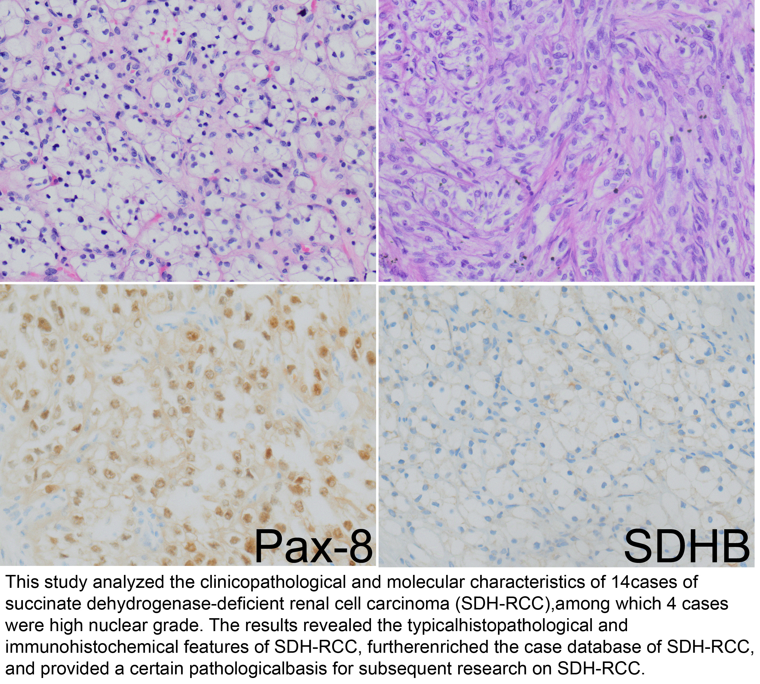
Results: Among the 14 patients, 10 were male and 4 were female. The age ranged from 43 to 78 years, with a median age of 52 years. 9 tumors were located in the left kidney and 5 in the right kidney. Tumor size ranged from 1.5 cm to 8.0 cm. Microscopically, the tumor cells exhibited diverse architectural patterns, including predominantly solid and nested growth, occasional microcystic and cystiic dilatation, and papillary architecture. The tumor cells had abundant cytoplasm, which was vacuolated or weakly eosinophilic. Immunohistochemical results showed that all 14 tumor cells were positive for vimentin and PAX-8, and negative for SDHB, CK7, CD117, and CA IX. The Ki-67 proliferation index ranged from 5% to 20%. Among the 14 patients, 1 was lost to follow-up, and 13 were followed up. Of the 13 followed patients, 12 were alive and 1 died.
Conclusion: SDH-deficient renal cell carcinoma is a relatively rare subtype of renal cell carcinoma. Patients with low nuclear grade are generally younger, whereas those with high nuclear grade are mostly middle-aged or elderly and prone to metastasis. Loss of SDHB expression by immunohistochemical staining is helpful for the diagnosis of this type of tumor.
References
[1] Wang G, & Rao P. (2018). Succinate Dehydrogenase-Deficient Renal Cell Carcinoma: A Short Review. Arch Pathol Lab Med, 142(10), 1284-1288. https://doi.org/10.5858/arpa.2017-0199-RS
[2] Dalla Pozza E, Dando I, Pacchiana R, Liboi E, Scupoli MT, Donadelli M, et al. (2020). Regulation of succinate dehydrogenase and role of succinate in cancer. Semin Cell Dev Biol, 98, 4-14. https://doi.org/10.1016/j.semcdb.2019.04.013
[3] Wu J, Wang Y, Yang L, Wang Y, Hu P, Fan L, et al. (2025). New Insights Into Succinate Dehydrogenase-Deficient Renal Cell Carcinoma. Arch Pathol Lab Med, 10.5858/arpa.2025-0233-OA. https://doi.org/10.5858/arpa.2025-0233-OA
[4] Moch H, Amin MB, Berney DM, Compérat EM, Gill AJ, Hartmann A, et al. (2022). The 2022 World Health Organization Classification of Tumours of the Urinary System and Male Genital Organs—Part A: Renal, Penile, and Testicular Tumours. European Urology, 82(5), 458-468. https://doi.org/https://doi.org/10.1016/j.eururo.2022.06.016
[5] Moch H, Cubilla AL, Humphrey PA, Reuter VE, & Ulbright TM. (2016). The 2016 WHO Classification of Tumours of the Urinary System and Male Genital Organs-Part A: Renal, Penile, and Testicular Tumours. Eur Urol, 70(1), 93-105. https://doi.org/10.1016/j.eururo.2016.02.029
[6] Gill AJ, Hes O, Papathomas T, Šedivcová M, Tan PH, Agaimy A, et al. (2014). Succinate dehydrogenase (SDH)-deficient renal carcinoma: a morphologically distinct entity: a clinicopathologic series of 36 tumors from 27 patients. Am J Surg Pathol, 38(12), 1588-1602. https://doi.org/10.1097/pas.0000000000000292
[7] Kamai T, Higashi S, Murakami S, Arai K, Namatame T, Kijima T, et al. (2021). Single nucleotide variants of succinate dehydrogenase A gene in renal cell carcinoma. Cancer Sci, 112(8), 3375-3387. https://doi.org/10.1111/cas.14977
[8] Sun X, Wang G, Huang Z, Li P, Yang B, Wang T, et al. (2023). Succinate Dehydrogenase Defects Giant Renal Cell Carcinoma. Urol Int, 107(8), 819-822. https://doi.org/10.1159/000531059
[9] Pan X, Wei Y, Sui X, Yin X, Zheng L, Zeng H, et al. (2024). [Succinate Dehydrogenase-Deficient Renal Cell Carcinoma: Clinicopathological Analysis of 11 Cases]. Sichuan Da Xue Xue Bao Yi Xue Ban, 55(5), 1099-1106. https://doi.org/10.12182/20240960101
[10] Wang XT, Wang X, Zhang RS, Cheng K, Xia QY, & Rao Q. (2022). [Succinate dehydrogenase-deficient renal cell carcinoma:a clinicopathological, ultrastructural and molecular analysis]. Zhonghua Bing Li Xue Za Zhi, 51(1), 12-16. https://doi.org/10.3760/cma.j.cn112151-20210823-00590
[11] Nezami BG, & MacLennan GT. (2025). Clear Cell Renal Cell Carcinoma: A Comprehensive Review of its Histopathology, Genetics, and Differential Diagnosis. Int J Surg Pathol, 33(2), 265-280. https://doi.org/10.1177/10668969241256111
[12] Henske EP, Cheng L, Hakimi AA, Choueiri TK, & Braun DA. (2023). Chromophobe renal cell carcinoma. Cancer Cell, 41(8), 1383-1388. https://doi.org/10.1016/j.ccell.2023.07.006
[13] Garje R, Elhag D, Yasin HA, Acharya L, Vaena D, & Dahmoush L. (2021). Comprehensive review of chromophobe renal cell carcinoma. Crit Rev Oncol Hematol, 160, 103287. https://doi.org/10.1016/j.critrevonc.2021.103287
[14] Williams GM, & Lynch DT. (2025). Renal Oncocytoma. In StatPearls. StatPearls Publishing Copyright © 2025, StatPearls Publishing LLC.
[15] Baranovska VV, Romanenko А, & Zakhartseva LM. (2020). Histological differential diagnostics of renal oncocytoma. Exp Oncol, 42(3), 233-237. https://doi.org/10.32471/exp-oncology.2312-8852.vol-42-no-3.14968
Type
Published
Data Availability Statement
The data used in this study are available from the corresponding author upon reasonable request.
Issue
Section
License
Copyright (c) 2026 Life Conflux

This work is licensed under a Creative Commons Attribution 4.0 International License.
How to Cite