The Mechanism of Platelet-Rich Plasma Added to Skin FlapPostoperative for the Influence of Skin Flap Survival
DOI:
https://doi.org/10.71321/0x94xp56Keywords:
free flap, Platelet-Rich Plasma Gel, Platelet-Rich Plasma, ultrasound contrast, VEGF, CD34Abstract
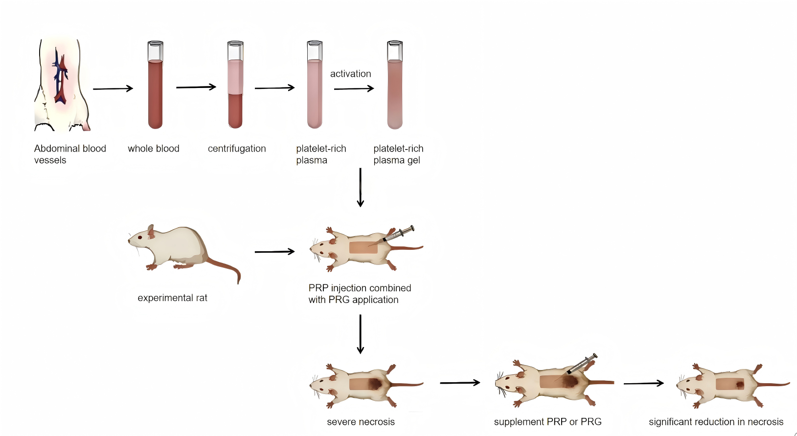
Background: This study investigated the effects of postoperative platelet-rich plasma (PRP) supplementation on skin flap survival in a rat model.
Methods: A random-pattern skin flap model was established in rats. During surgery, PRP injection combined with platelet-rich gel (PRG) application was performed. On postoperative day 3, PRP or PRG was supplemented into the flap via uniform point injections. Necrotic area was recorded at postoperative day 7. Flap defect length and terminal edema severity were assessed by contrast-enhanced ultrasonography. Tissue specimens were collected for histopathological and immunohistochemical analysis. Relative expression levels of vascular endothelial growth factor (VEGF), platelet-derived growth factor (PDGF), and cluster of differentiation 34 (CD34) were measured.
Results: The group receiving intraoperative PRP injection combined with PRG application, followed by postoperative PRG supplementation, showed significantly shorter tissue defect lengths and less terminal tissue edema compared to other groups (P<0.01). Relative expression levels of VEGF, PDGF, and CD34 were significantly higher than those in other groups (P<0.01).
Conclusion: Postoperative PRP and PRG supplementation reduced flap defect length and edema severity, increased growth factor expression, and improved flap survival. PRG supplementation produced the most pronounced benefits.
References
[1] Ershadifar, S., Colback, A., Basmaci, U. N., Wilson, M., Birkeland, A. C., and Silverman, D. A. Predictors of Donor-Site Wound Complications Following Fibula Free Flap Reconstruction. OTO Open. 2025;9(2):e70126. https://doi.org/10.1002/oto2.70126
[2] Yang, J., Qin, X., Hou, L., and Liu, Y. Risk prediction models for complications after flap repair surgery: a systematic review and meta-analysis. BMC Surgery. 2025;25(1):398. https://doi.org/10.1186/s12893-025-03072-8
[3] Marx, R. E. Platelet-rich plasma: evidence to support its use. Journal of Oral and Maxillofacial Surgery. 2004;62(4):489–496. https://doi.org/10.1016/j.joms.2003.12.003
[4] Everts, P., Onishi, K., Jayaram, P., Lana, J. F., and Mautner, K. Platelet-Rich Plasma: New Performance Understandings and Therapeutic Considerations in 2020. International Journal of Molecular Sciences. 2020;21(20):7794. https://doi.org/10.3390/ijms21207794
[5] Yang, D., and Morris, S. F. Comparison of two different delay procedures in a rat skin flap model. Plastic and Reconstructive Surgery. 1998;102(5):1591–1597. https://doi.org/10.1097/00006534-199810000-00039
[6] Zhang, D., Jin, C., Han, T., Chen, J., Ali Raza, M., Li, B., et al. Sinomenine promotes flap survival by upregulating eNOS and eNOS-mediated autophagy via PI3K/AKT pathway. International Immunopharmacology. 2023;116:109752. https://doi.org/10.1016/j.intimp.2023.109752
[7] Harder, Y., Amon, M., Erni, D., and Menger, M. D. Evolution of ischemic tissue injury in a random pattern flap: a new mouse model using intravital microscopy. Journal of Surgical Research. 2004;121(2):197–205. https://doi.org/10.1016/j.jss.2004.03.026
[8] Milton, S. H. Pedicled skin-flaps: the fallacy of the length: width ratio. British Journal of Surgery. 1970;57(7):502–508. https://doi.org/10.1002/bjs.1800570705
[9] Lee, J. H., You, H. J., Lee, T. Y., and Kang, H. J. Current Status of Experimental Animal Skin Flap Models: Ischemic Preconditioning and Molecular Factors. International Journal of Molecular Sciences. 2022;23(9):5234. https://doi.org/10.3390/ijms23095234
[10] Landén, N. X., Li, D., and Ståhle, M. Transition from inflammation to proliferation: a critical step during wound healing. Cellular and Molecular Life Sciences. 2016;73(20):3861–3885. https://doi.org/10.1007/s00018-016-2268-0
[11] Eming, S. A., Martin, P., and Tomic-Canic, M. Wound repair and regeneration: mechanisms, signaling, and translation. Science Translational Medicine. 2014;6(265):265sr6. https://doi.org/10.1126/scitranslmed.3009337
[12] Collins, T., Alexander, D., and Barkatali, B. Platelet-rich plasma: a narrative review. EFORT Open Reviews. 2021;6(4):225–235. https://doi.org/10.1302/2058-5241.6.200017
[13] Boswell, S. G., Cole, B. J., Sundman, E. A., Karas, V., and Fortier, L. A. Platelet-rich plasma: a milieu of bioactive factors. Arthroscopy. 2012;28(3):429–439. https://doi.org/10.1016/j.arthro.2011.10.018
[14] Akbarzadeh, S., McKenzie, M. B., Rahman, M. M., and Cleland, H. Allogeneic Platelet-Rich Plasma: Is It Safe and Effective for Wound Repair? European Surgical Research. 2021;62(1):1–9. https://doi.org/10.1159/000514223
[15] Ferrara, N., and Kerbel, R. S. Angiogenesis as a therapeutic target. Nature. 2005;438(7070):967–974. https://doi.org/10.1038/nature04483
[16] Carmeliet, P., and Jain, R. K. Molecular mechanisms and clinical applications of angiogenesis. Nature. 2011;473(7347):298–307. https://doi.org/10.1038/nature10144
[17] Vourtsis, S. A., Spyriounis, P. K., Agrogiannis, G. D., Ionac, M., and Papalois, A. E. VEGF application on rat skin flap survival. Journal of Investigative Surgery. 2012;25(1):14–19. https://doi.org/10.3109/08941939.2011.593693
[18] Kryger, Z., Zhang, F., Dogan, T., Cheng, C., Lineaweaver, W. C., and Buncke, H. J. The effects of VEGF on survival of a random flap in the rat: examination of various routes of administration. British Journal of Plastic Surgery. 2000;53(3):234–239. https://doi.org/10.1054/bjps.1999.3315
[19] Shang, Y., Xie, X., Luo, Y., Nie, F., Luo, Y., Jing, X., et al. Safety findings after intravenous administration of sulfur hexafluoride microbubbles to 463,434 examinations at 24 centers. European Radiology. 2023;33(2):988–995. https://doi.org/10.1007/s00330-022-09108-4
[20] Newsome, I. G., and Dayton, P. A. Acoustic Angiography: Superharmonic Contrast-Enhanced Ultrasound Imaging for Noninvasive Visualization of Microvasculature. Methods in Molecular Biology. 2022;2393:641–655. https://doi.org/10.1007/978-1-0716-1803-5_34
[21] Lv, K., Zhai, H., Jiang, Y., Liang, P., Xu, H. X., Du, L., et al. Prospective assessment of diagnostic efficacy and safety of SonazoidTM and SonoVue® ultrasound contrast agents in patients with focal liver lesions. Abdominal Radiology. 2021;46(10):4647–4659. https://doi.org/10.1007/s00261-021-03010-1
Type
Published
Data Availability Statement
All data needed to evaluate the conclusions in the paper are present in the paper or the Supplementary Materials. Additional data related to this paper may be requested from the authors.
Issue
Section
License
Copyright (c) 2026 Life Conflux

This work is licensed under a Creative Commons Attribution 4.0 International License.
How to Cite